
Free preview

Medical conditions affecting the endocrine system

DN 222 · Medical nursing (III) — Diploma Nursing - Direct

Learning objectives

  • Applied anatomy and physiology of the endocrine system
  • Acromegaly
  • Dwarfism
  • Addison's disease
  • Cushing's syndrome
  • Pheochromocytoma
  • Hyperaldesteronism

Introductory study notes

Applied anatomy and physiology of the endocrine system

Introduction

The endocrine system is made up of ductless glands that secrete hormones directly into the blood. Hormones travel to target cells that have specific receptors and regulate growth, metabolism, fluid and electrolyte balance, reproduction and the stress response. It works with the nervous system to maintain homeostasis; the nervous system acts in milliseconds, the endocrine system more slowly but for longer.

Types of hormones

  • Steroid hormones – made from cholesterol, lipid-soluble, act on receptors inside the cell (cortisol, aldosterone, oestrogen, testosterone).
  • Peptide/protein hormones – water-soluble, act on cell-membrane receptors (insulin, GH, ADH).
  • Amine hormones – derived from amino acids (adrenaline, thyroxine, melatonin).

Control of hormone secretion

Most hormones are controlled by negative feedback: a rising hormone level switches off its own stimulus (e.g. high thyroxine suppresses TRH and TSH). Positive feedback is rare – e.g. oxytocin during labour increases contractions, which release more oxytocin.

Main glands and hormones

Gland

Hormones and actions

Hypothalamus

Releasing/inhibiting hormones (TRH, CRH, GnRH, GHRH, dopamine) that control the pituitary; links nervous and endocrine systems

Anterior pituitary ("master gland")

GH (growth), TSH (stimulates thyroid), ACTH (stimulates adrenal cortex), FSH and LH (gonads), prolactin (milk production)

Posterior pituitary

Stores and releases ADH (water reabsorption in collecting ducts) and oxytocin (uterine contraction, milk let-down) made in the hypothalamus

Thyroid

T3 and T4 (need iodine; raise metabolic rate, growth and brain development); calcitonin (lowers blood calcium)

Parathyroids

Parathyroid hormone – raises blood calcium (bone resorption, kidney reabsorption, activates vitamin D)

Adrenal cortex

Zona glomerulosa – aldosterone (Na⁺ and water retention, K⁺ excretion); zona fasciculata – cortisol (raises glucose, stress response, anti-inflammatory); zona reticularis – androgens

Adrenal medulla

Adrenaline and noradrenaline – fight-or-flight

Pancreas (islets of Langerhans)

β-cells – insulin (lowers blood glucose); α-cells – glucagon (raises blood glucose); δ-cells – somatostatin

Gonads

Ovaries – oestrogen, progesterone; testes – testosterone

Pineal gland

Melatonin – sleep–wake cycle

Thymus

Thymosin – maturation of T-lymphocytes

Nursing relevance

Nurses monitor vital signs, fluid balance, weight, blood glucose and electrolytes; give hormone replacement at the correct times; and teach patients never to stop long-term steroids abruptly.
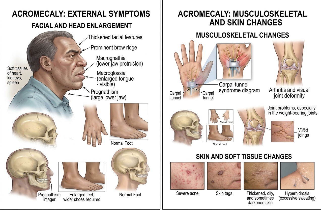
Acromegaly

Diagram showing the symptoms of acromegaly.

Acromegaly.

Acromegaly is a rare disorder of excessive bone and soft tissue growth due to elevated levels of growth hormone.
In young children, prior the completed fusion and growth of bones, excessive growth hormone can cause a similar condition called gigantism.

Causes.

In most cases, acromegaly is caused by over-secretion of growth hormone (GH) produced by a benign tumor of the pituitary gland.
The pituitary gland is a small gland located at the base of the brain that produces many hormones.
In a small number of cases, malignant tumors of other organs (pancreas, adrenal, lung) may be the source of excess GH.

Risk factors.

Some rare cases of acromegaly are hereditary.

The average age of diagnosis is 40-45 years old.

Symptoms.

Symptoms usually develop very slowly over time.

Acromegaly can cause serious complications and premature death if not treated.

In children, excess GH production causes elongation of the bones and associated soft tissue swelling.
If not treated, children with this disorder can grow ta height of 7-8 feet.

Symptoms and complications in adults may include the following:

  1. Abnormally large growth and deformity of the:

  2. Hands (rings no longer fit)

  3. Feet (need a bigger size shoe)

  4. Face (protrusion of brow and lower jaw)

  5. Jaw (teeth do not line up correctly when the mouth is closed)

  6. Lips

  7. Tongue

  8. Carpal tunnel syndrome

  9. Skin changes, such as:

  10. Thickened, oily, and sometimes darkened skin

  11. Severe acne

  12. Excessive sweating and offensive body order due tenlargement of the sweat glands

  13. Deepening voice due tenlarged sinuses, vocal cords, and soft tissues of the throat

  14. Fatigue and weakness in legs and arms

  15. Sleep apnea

  16. Arthritis and other joint problems especially in the jaw

  17. Hypothyroidism

  18. Enlargement of the liver, kidneys, spleen, heart, and/or other internal organs, which can lead to:

a)Diabetes

b)High blood pressure

c)Cardiovascular disease

In women:

a)Irregular menstrual cycles

b)Galactorrhea (abnormal production of breast milk) in 50% of cases

n men:

In about 50% of cases, impotence

Diagnosis.

Blood tests will be done to measure the level of insulin-like growth factor (IGF-I), growth hormone releasing hormone (GHRH), and other pituitary hormones.

A glucose tolerance test may also be given to see if the GH level drops—it will not drop in cases of acromegaly.

If these tests confirm acromegaly, the following may be done to locate the tumor that is causing the disorder:

Head CT Scan a type of x-ray that uses a computer to make pictures of the inside of the brain and surrounding structures

MRI Scan

Treatment.

Reduce production of GH to normal levels

Stop and reverse the symptoms caused by over-secretion of GH

Correct other endocrine abnormalities (thyroid, adrenal, sex organs)

Reduce the tumor size

Treatment may include:

Surgery.

Surgical removal of the pituitary tumor, or other tumor, that is believed to be causing acromegaly may be done.
In most cases, this is the preferred treatment.

Radiosurgery

Radiosurgery is the use of highly focused external beams of radiation to shrink the tumor.
It is used most often in patients who do not respond to conventional surgery or medications.

Radiation Therapy

Radiation therapy is used in combination with either medical and/or surgical treatment.

Medication

Drugs may be given to reduce the level of GH secretion from the pituitary gland.
These include:

Cabergoline given orally

Pergolide given orally

Bromocriptine may be given before surgery to shrink tumor

Octreotide given by injections

Pegvisomant given by injections for patients not responding to other forms of treatment

Medications must often be combined with other therapies to treat larger tumors affecting surrounding structures.

Prevention.

There are guidelines for preventing acromegaly.
Early diagnosis and treatment, however, will help prevent serious complications, some of which are irreversible.

Pheochromocytoma

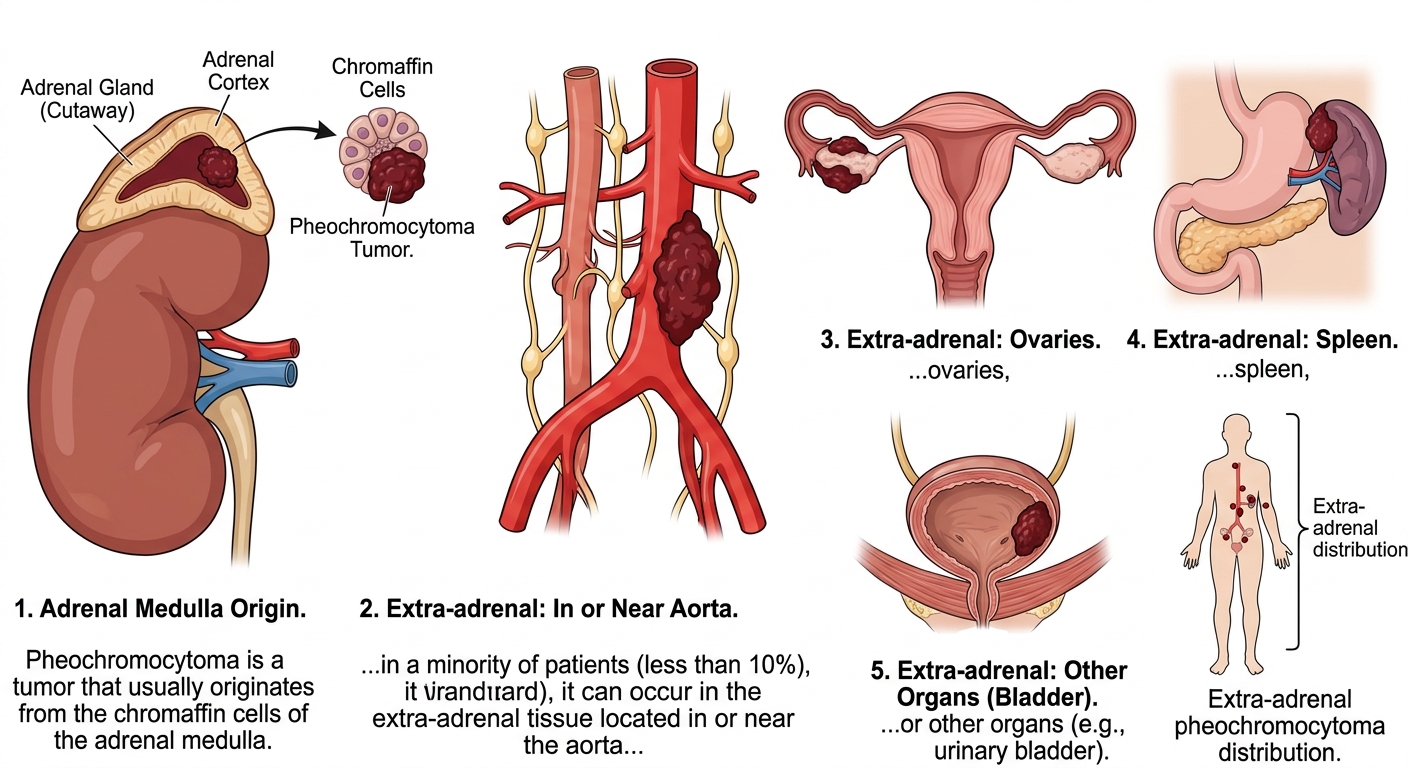
Diagram showing what Pheochromocytoma.

Pheochromocytoma.

Pheochromocytoma is a tumor that usually originates from the chromaffin cells of the adrenalmedulla; in a minority of patients (less than 10%), it can occur in the extra-adrenal tissue located in or near the aorta, ovaries, spleen, or other organs

Pheochromocytoma can occur at any age, but its peak incidence is from ages 40 to 50; it affects men and women equally and has a genetic association.

Signs and symptoms.

  1. The type and severity of signs and symptoms depends on the relative proportions of epinephrine and norepinephrine secreted; approximately 8% of patients are completely asymptomatic

  2. In patients with hypertension, the typical triad of signs and symptoms is headache, diaphoresis, and palpitations; hypertension may be intermittent or persistent

  3. Other signs and symptoms may include tremor, flushing, anxiety, and hyperglycemia

  4. The paroxysmal form of pheochromocytoma is typically characterized by acute, unpredictable attacks that last from seconds to several hours, with symptoms typically beginning abruptly and subsiding slowly; the patient becomes extremely anxious, tremulous, and weak and may have such other symptoms as tachycardia, nausea, vomiting, diarrhea, abdominal pain, and a feeling of impending doom

  5. Severe hypertension can cause serious, life-threatening complications, such as cardiac arrhythmias, dissecting aneurysm, stroke, and acute renal failur

  6. Postural hypotension occurs in 70% of patients with untreated pheochromocytoma

Diagnosis and treatment.

  1. Imaging studies, such as CT scanning, MRI, and ultrasonography, can localize the pheochromocytomas and determine whether the patient has more than one tumor

  2. Pheochromocytoma is suspected in the patient with signs of sympathetic nervous system over activity, such as hypertension, headache, excessive sweating, hyper metabolism, and hyperglycemia

  3. Increased urine and plasma levels of catecholamines and metanephrine, a catecholamine metabolite, are the most direct and conclusive tests for over activity of the adrenal medulla

  4. If urine and plasma tests of catecholamines are inconclusive, a clonidine suppression test can identify excess catecholamine release that bypasses normal storage and release mechanisms, a characteristic of pheochromocytoma

  5. Initial treatment aims to reduce blood pressure to normal levels; drugs used include alpha-adrenergic, beta-adreergic, and calcium channel blockers and smooth muscle relaxants

  6. If other medications don’t control blood pressure, catecholamine synthesis inhibitors may be used

  7. The definitive treatment for Pheochromocytoma is surgical removal of the tumor; it’s usually accompanied by adrenalectomy

  8. A patient who has undergone bilateral adrenalectomy requires corticosteroid replacement

  9. Postoperatively, the patient’s blood pressure and catecholamine levels should return to normal; if not, he should be evaluated for retention of Pheochromocytoma tissue

Specific nursing interventions/specific nursing care,

1.      Monitor the patient’s blood pressure, and administer antihypertensive medications as ordered

2.      Prepare the patient for surgery; ensure that he’s well hydrated to prevent hypotension

3.      Manipulation of the tumor during surgical excision may cause the release of stored epinephrine and norepinephrine, with marked increases in blood pressure and heart rate changes; monitor the patient closely

4.      Administer corticosteroids as ordered to prevent adrenal insufficiency

5.      Monitor for hypoglycemia

6.      Collect blood and urine samples for catecholamine and metabolite testing to evaluate complete tumor removal

7.      Teach the patient and family about the disease, including treatments and signs of recurrence; teach them how to monitor the patient’s blood pressure at home

8. Provide emotional support to the patient and family, who may fear repeated attacks

More free previews in Medical nursing (III)